Thursday, November 30, 2017

World AIDS Day 2017

World Aids Day

World AIDS Day takes place on December 1st every year, providing an opportunity for people globally to unite in the fight against HIV, show support for people living with HIV, and to commemorate those who died from AIDS-related illness.

World AIDS Day was the 1st global health day, founded in 1988, with the Red Ribbon becoming an internationally recognised symbol of HIV and AIDS awareness; created by the group Visual AIDS in 1991. The red ribbon is worn by people throughout the year in support of people living with HIV and in remembrance of those who have died, but on World AIDS Day the red ribbon highlights the solidarity for people living with HIV and is worn as a simple yet powerful way to challenge stigma and prejudice.

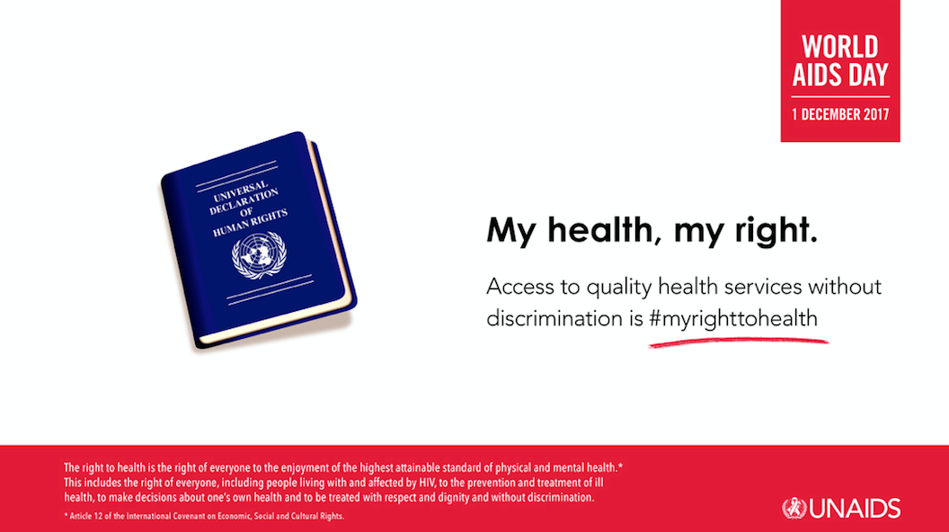
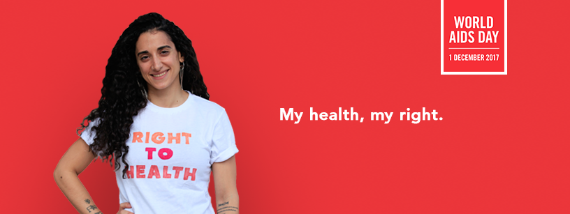
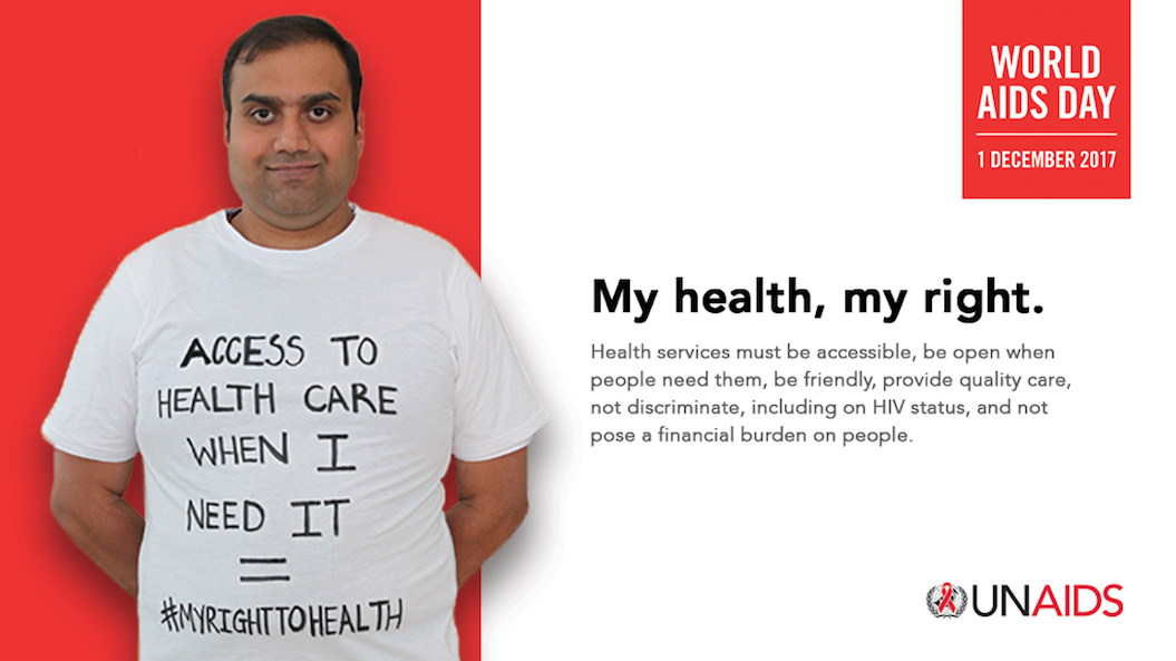
This year there are many campaigns for World AIDS Day 2017 (#WAD2017). In the United States of America, this years theme from the U.S Agency for international Development (USAID) is “Increasing Impact through Transparency, Accountability, and Partnerships”; reflecting on the impact of global partnerships.  In the United Kingdom the theme from the British HIV Association (BHIVA) is “HIV: Towards Zero” (#HIVZero) to celebrate the progress and achievements made in HIV diagnosis and management, whilst highlighting the challenges that remain. The South African National AIDS Council (SANAC) has adopted the global UNAIDS theme of “Right to Health” (#myrighttohealth); the right of everyone to the enjoyment of the highest attainable standards of physical and mental health. This includes people living with and affected by HIV, to the prevention and treatment of ill health, to make decisions about ones own health and to be treated with respect and dignity and without discrimination. Almost all of the Sustainable Development Goals are linked in some way to health, so achieving the Sustainable Development Goals (which includes ending the AIDS epidemic) depends on ensuring the “Right to Health”.

World Aids Day

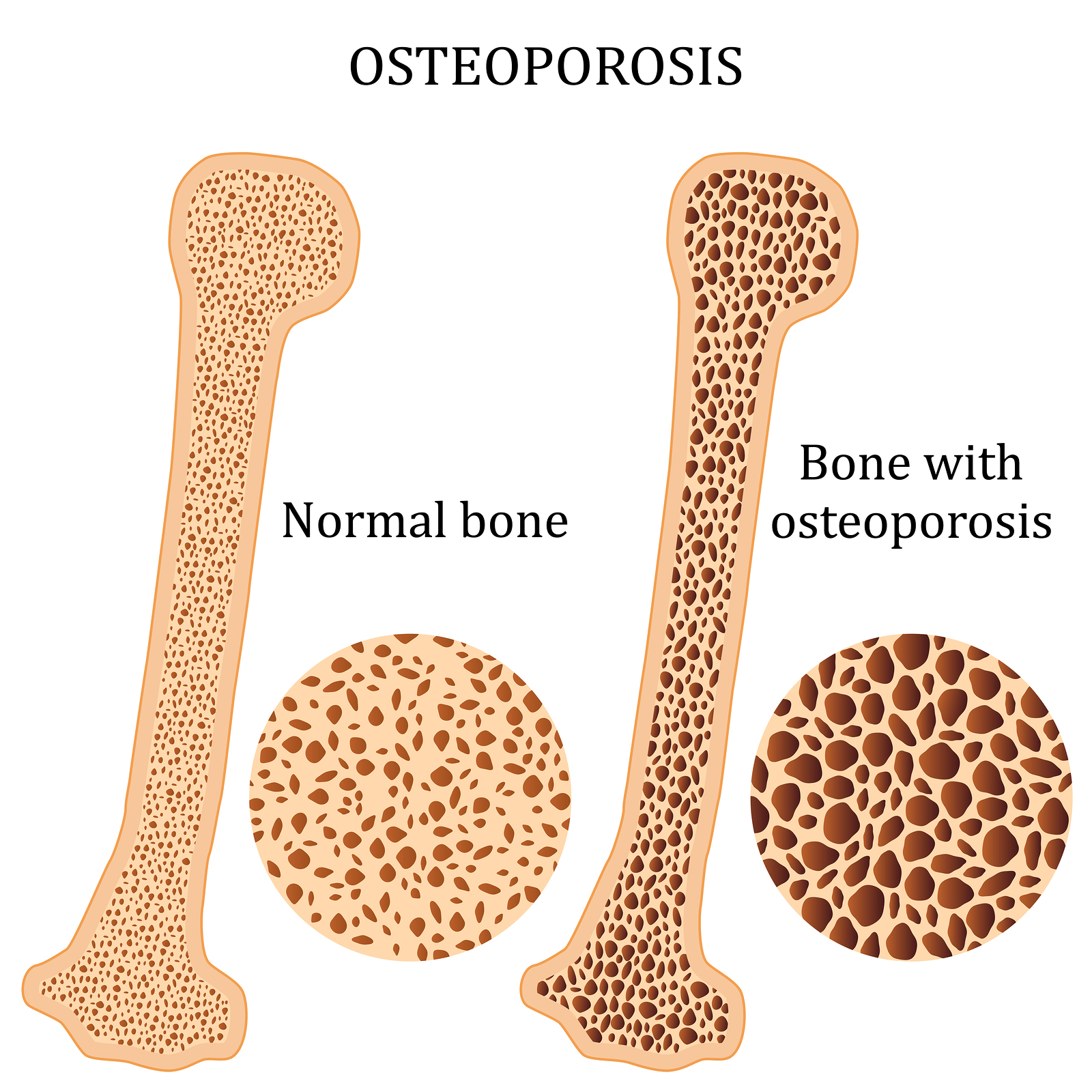
Good health is a prerequisite for progress on ending AIDS. Ensuring healthy lives and promoting well-being for all at all ages, including people living with or at risk of HIV, is essential to sustainable development. In my previous blogs I have spoken about the role that Physiotherapists play in supporting people living with HIV. Increasing service integration in a way that responds to individuals’ needs, can lead the way in reshaping efficient, accessible and equitable health services for HIV and beyond – and this should include Physiotherapy and rehabilitation. The World Health Organisations “Rehabilitation 2030: a call to action” highlights the increasing unmet needs to people with long term health conditions globally, experiencing disability and requiring rehabilitation. So for World AIDS Day 2017, I want to champion the role of Physiotherapists in supporting people living with HIV, to live healthy lives and promote well-being for all at all ages. Physiotherapy and Rehabilitation fits into this year’s theme of “Right to Health”, because it is a right to access Physiotherapy for people living with HIV who experience disability, cannot engage in exercise, are frail or have unmet rehabilitation needs.

World AIDS Day 2017 is a chance to celebrate successes and highlight remaining challenges, but it must also be a time for reflection and remembrance to all those we have lost; as demonstrated in this recent article from the Guardian about the book “The Ward”, which has an exhibition in London until 3rd December. This reflection should allow us to feel proud in how much HIV has changed, how “we live in such different times”, and how we will never forget those we loved, those we cared for, those we lost.

So this World AIDS Day, wear your red ribbon with pride, passion, solidarity, support, anger, love, compassion, in memory, or in any way you that feel right for you.

World Aids Day

Clinical implications from daily physiotherapy of 131 acute hamstring injuries & their association with rehab

Young woman with pulled hamstring. Hamstring pain after sport playing.

The aim of this study was to investigate the association of daily clinical measures and the progression of rehabilitation and perceived running effort. A cohort of 131 athletes with an MRI-confirmed acute hamstring injury underwent a standardised criteria-based rehabilitation protocol. Descriptive and inferential statistics were used to investigate the association between daily clinical subjective and objective measures and both the progression of rehabilitation and perceived running effort. These measures included different strength, palpation, flexibility and functional tests. Inter-rater and intrarater reliability and minimal detectable change were established for the clinical measures of strength and flexibility by examining measures taken on consecutive days for the uninjured leg.

The progression of the daily measures was seen to be non-linear and varied according to the measure. Intra-rater reliability for the strength and flexibility measures were excellent (95% CI ≥0.85 for all measures). Strength (in the outer range position) and flexibility (in maximum hip flexion with active knee extension (MHFAKE) in supine) were best associated with rehabilitation progression and perceived running effort. Additionally, length of pain on palpation was usefully associated with rehabilitation progression. At lower perceived running effort there was a large variation in actual running speed. Daily physical measures of palpation pain, outer range strength, MHFAKE and reported pain during daily activity are useful to inform the progression of rehabilitation.

Effect of aerobic exercise on hippocampal volume in humans: A systematic review and meta-analysis.

Neurons, marked by fluorescence

Hippocampal volume increase in response to aerobic exercise has been consistently observed in animal models. However, the evidence from human studies is equivocal. The authors undertook a systematic review to identify all controlled trials examining the effect of aerobic exercise on the hippocampal volumes in humans, and applied meta-analytic techniques to determine if aerobic exercise resulted in volumetric increases. They also sought to establish how volume changes differed in relation to unilateral measures of left/right hippocampal volume, and across the lifespan.

A systematic search identified 4398 articles, of which 14 were eligible for inclusion in the primary analysis. A random-effects meta-analysis showed no significant effect of aerobic exercise on total hippocampal volume across the 737 participants. However, aerobic exercise had significant positive effects on left hippocampal volume in comparison to control conditions. Post-hoc analyses indicated effects were driven through exercise preventing the volumetric decreases which occur over time.

These results provide meta-analytic evidence for exercise-induced volumetric retention in the left hippocampus. Aerobic exercise interventions may be useful for preventing age-related hippocampal deterioration and maintaining neuronal health.

Why Aren't We Turning Off the Light Switch More?


Remember the Clapper?  “Catchy jingle” is how Morgan Freeman described it in Bruce Almighty.  A simple device applied to a light switch that is designed to turn the light on or off when a clapping of the hands happens.  Super popular in the 90’s. “Clap on…Clap off….the Clapper”. Got me thinking after I saw that the clapper was still available for purchase…what if…as rehabilitation professionals we were able to turn off a client’s symptoms during their visit instead of producing them?  What if…we were able to do that and then show the client how to reproduce that themselves?  Now I’m not talking about avoiding pain.  I’m not even talking about talking it easy on our clients in rehab sessions by not having them work hard or achieve muscular fatigue during exercise.  But rather, I’m suggesting, that as part of an assessment tool, we try to determine if the use of forces around the body with our hand, education, postural changes, belts, etc, can turn the symptom ‘off’ instead of ‘on’.  What information would that give us if we could do that?  With all the social media ‘disruptors’ and challengers out there (nothing wrong with it), it seems like we’re being convinced that we haven’t gotten ANYONE better in the past 50 years!  Successes that we have seen appear to be merely a fog curtain of Hawthorne effects and placebos. Our biggest challenge in healthcare is NOT the mechanisms of how we get people better.  Not even the application to get to the mechanism.  Our biggest challenge is the clinical thought process of why we do what we do!  In 17 years as a physio, I’ve gone through the doldrums of success and failure.  I’ve convinced myself that I was top of the food chain, and sweated the disbelief of letting my patients down of not achieving their goals. 
One of my biggest epiphany however came from the realization that I could get just as much (if not more information) about my patient from attempting to eliminate their symptoms alongside trying to stimulate them.  @Keyclinicalskills refers to this as “Light switch off.”  By turning my attention away from “does this hurt,” “ooooo this feels restricted” and the ever loved “this end-feel feels tight,” and focusing on “do your symptoms change when I do this?”  Recently published studies have even demonstrated dramatic changes in cerebral processing are being seen with fMRI just by changing the words we use and the focus we place on movement (found here).  I have found several things have happened when I attempt light switch off testing and treatment.  1) I automatically engage the patient! Positively! Establishing a therapeutic alliance is becoming more documented in the literature and is defined as the working rapport or positive social connection between the patient and the therapist 
 (found here).  2) Buy in from the patient.  We have established our profession as the “Physical Torturists” or “Personal Terrorist.”  All lovely titles! From the outset of rehabilitation in the physical therapy setting, some people are automatically expecting pain when they come in for their visit.  “Just do what you have to do” resonates throughout clinics across the globe as we have convinced people the only way to get better is through trial by fire and pain! Why is this?  Are we finding justification through experience and research that lightly suggests that painful treatment are better than non?  Have we given up on the possibility that turning off pain could lead to normalcy? 
Now this suggestion, while logical, is a huge uphill battle.  I cannot remember any aspect of learning how to look for pain free objective signs in PT.  My education, both during schooling, and in most of my 574 hours of post-graduate continuing education since has been focused around pain producing special tests. 
Problem?  Chad Cook and Eric Hedeges would argue yes! 
Chad and Eric have authored 2 different documents that are leading our profession to question what we are even looking at with our ‘special tests’.  One is Orthopedic Special Tests Volume 2 and the other is an opinion piece published in JOPST earlier this year (2017).  The over-simplified conclusion?  We have fooled ourselves with biased research and a misunderstanding of previously conducted studies that have yielded and abundance of unreliable special test…that aren’t that special!
So back to my early statements.  What kind of value could be place on performing tests that were designed to eliminated symptoms instead of producing them?  I have established a habit over the last several years of attempting forms of pain-eliminating tests first to determine results, then returning to the pain-producing testing after to compared notes. 
Something interesting that I have found from symptom-eliminating testing is the response from the patient.  Case in point, imagine the painful knee patient who has already been told that they have advanced arthritis in their knee and that portions of their meniscus are torn after MRI.  Often, they are unsure as to why they are in therapy because their condition cannot change and their beliefs have been established from a consult from a healthcare professional who has not educated them on pain or the research findings that knee OA does not equal pain and dysfunction for every person.  If I perform painful special tests on them in an attempt to produce pain or limitations first, I often justify to them their beliefs of the problem or issue.  Now, what if I attempt the opposite.  What if Mrs. Jones is a responder to improved pain-free ROM and function with simple forces applied across her joint line while she performs that actions that she describes at limiting?  “What happened to my OA?” is the most common response I get. “Exactly my point. Nothing happened to your OA Mrs. Jones.  But now we know that your body has the capability to operative without that limitation.  So now we just have to figure out how to make that change permanent.”  Moving toward a complimentary team effort with my patient about what means the most to them and away from fully passive treatments has made it easier to establish patient empowerment and control over their symptoms.  People feel listened to. 2) If symptoms can be altered, and preferable abolished, it automatically begins that question of “If I can move without that pain or restriction now, what happened to my “X” (insert OA, tear, degeneration, posture, etc) diagnosis?”.  The transition from a diagnosis to Pain Neuro Science (PNE) happens seamlessly. Beginning to understand that the vast majority of symptoms that we see clinically in an outpatient setting fall under the category of “top down”.
Imagine a world where you were above to move and function without pain?!?  Yeah…it’s called normal!

It may be easily argued that the Therapeutic Alliance has already begun in this scenario.  The patient would likely “buy in” to the treatment approach quickly with a positive response.  Especially compared to the more traditional approach of “light switch on” special testing and treatment. But “light switch on” not only does have value proven in the literature (see cross over leg raise example here), but working with pain can have its advantages as well.  I often have my clients focus on the pain that they are having and determine what happens to their pain when they move or exercise.  “Ok.  Let’s see what happens to your pain or limitation if you do more repetitions?  Is it getting worse, better, or staying the same the more you do?  I classically get even more information after a bout of painful exercise or movements…”now let’s try that again and tell me what has happened to that pain you were reporting earlier.”
Unfortunately, while there have been countless observations in the literature of these types of treatments (see here), to my knowledge there have not been any performed directly related to specific conditions.  But do we really need specific research to perform “light switch off” testing?  Can we really infer that a sub-group of responders would give any value over an N of 1? 
Perhaps we have given the painful approach too much credence over time.  Have we relied on special tests and painful treatments to define who we are as a profession?  Maybe. Is it time for a paradigm shift in how we conduct an exam and treatment and start including pain-free testing and treatment?  Maybe it’s time to consider both pain free manual therapy and pain free exercise.  And if we can do that, then LOAD IT!  Load it heavy, and often, with current suggestions from Prof. Jill Cook among others. 
Thanks for reading.  Now that you're done, turn off the light please!
lead instructor The Eclectic Approach to Modern Motor Control


Want an approach that enhances your existing evaluation and treatment? No commercial model gives you THE answer. You need an approach that blends the modern with the old school. Live cases, webinars, lectures, Q&A, hundreds of techniques and more! Check out Modern Manual Therapy!

Keeping it Eclectic...

Cross-education of muscular strength following unilateral resistance training: a meta-analysis.

Long Receding Row Of Kettlebell Weights In A Gym

Cross-education (CE) of strength is a well-known phenomenon whereby exercise of one limb can induce strength gains in the contralateral untrained limb. The only available meta-analyses on CE, which date back to a decade ago, estimated a modest 7.8% increase in contralateral strength following unilateral training. However, in recent years new evidences have outlined larger contralateral gains, which deserve to be systematically evaluated. Therefore, the aim of this meta-analysis was to appraise current data on CE and determine its overall magnitude of effect.

Five databases were searched from inception to December 2016. All randomized controlled trials focusing on unilateral resistance training were carefully checked by two reviewers who also assessed the eligibility of the identified trials and extracted data independently. The risk of bias was assessed using the Cochrane Risk-of-Bias tool.

Thirty-one studies entered the meta-analysis. Data from 785 subjects were pooled and subgroup analyses by body region (upper/lower limb) and type of training (isometric/concentric/eccentric/isotonic-dynamic) were performed. The pooled estimate of CE was a significant 11.9% contralateral increase (95% CI 9.1-14.8; p < 0.00001; upper limb: + 9.4%, p < 0.00001; lower limb: + 16.4%, p < 0.00001). Significant CE effects were induced by isometric (8.2%; p = 0.0003), concentric (11.3%; p < 0.00001), eccentric (17.7%; p = 0.003) and isotonic-dynamic training (15.9%; p < 0.00001), although a high risk of bias was detected across the studies. Unilateral resistance training induces significant contraction type-dependent gains in the contralateral untrained limb. Methodological issues in the included studies are outlined to provide guidance for a reliable quantification of CE in future studies.

Wednesday, November 29, 2017

Trajectories of the relationships of physical activity with body composition in older men: the MrOS study.

Fitness at home: free weights, yoga mat, mobile phone app. Dumbb

Excess adiposity gains and significant lean mass loss may be risk factors for chronic disease in old age. Long-term patterns of change in physical activity (PA) and their influence on body composition decline during aging has not been characterized. The authors evaluated the interrelationships of PA and body composition at the outset and over longitudinal follow-up to changes in older men.

Self-reported PA by the Physical Activity Scale for the Elderly (PASE), clinic body weight, and whole-body lean mass (LM) and fat mass, by dual-energy x-ray absorptiometry (DXA), were assessed in 5964 community-dwelling men aged ≥65 years at baseline (2000-2002) and at two subsequent clinic visits up until March 2009 (an average 4.6 and 6.9 years later). Group-based trajectory modeling (GBTM) identified patterns of change in PA and body composition variables. Relationships of PA and body composition changes were then assessed.

GBTM identified three discrete trajectory patterns, all with declining PA, associated primarily with initial PA levelshigh-activity (7.2% of men), moderate-activity (50.0%), and low-activity (42.8%). In separate models, GBTM identified eight discrete total weight change groups, five fat mass change groups, and six LM change groups. Joint trajectory modeling by PA and body composition group illustrated significant declines in total weight and LM, whereas fat mass levels were relatively unchanged among high-activity and low-activity-declining groups, and significantly increased in the moderate-activity-declining group.

Although patterns of change in PA and body composition were identified, groups were primarily differentiated by initial PA or body composition rather than by distinct trajectories of change in these variables.

Fixing Knee Pain in Runners

Running Athlete Feeling Pain Because Of Injured Knee

Collapsing

In my last post, we discussed the idea of a collapsing runner. This is someone who either shows a Trendelenberg in the stance phase of gait, or collapses inward at the knee through hip internal rotation and adduction. This is common with runners experiencing knee pain.

Many times I find the typical prescription is hip strengthening. It makes perfect sense. The hip abductors and external rotators do not possess enough strength to control the collapse. Many runners will see improved pain levels with this plan, but it is also important to realize that many runners may have a reoccurrence of the pain at a later time. Is it possible that one of the reasons your runner improved is partially due to the time they likely spent reducing their running volume while in rehab?

Here’s the thing. Just because you improve a runner’s hip strength does not mean his/her running gait will change.²  If running brought on the pain and we’re not fixing the running mechanics, then we’re addressing only half the problem!

With that said if you’re not fixing the strength deficits, your ability to fix the running likely may also not be successful.  It is vitally important to know whether your fix needs to be aimed at mobility, motor control, strength, or a running technique impairment.  I use the ACE Running Movement and Stability Screen then perform a running gait analysis  to determine what area(s) I should be addressing.

Once there is adequate mobility, strength, and pain is under control, it is imperative that we move a step past just strengthening and perform gait retraining. There are many ways to accomplish this.  Just a few options are:

1. Biofeedback: I use Trace 3D motion capture, but you can also use a mirror or another form of biofeedback to cue the patient
2. Provide verbal cues, such as, “pretend there are flashlights on your knees, and point them straight ahead”
3. Manipulate Cadence

Cadence

We’re going to focus on manipulating cadence. Many runners demonstrate a slower than ideal cadence. If you’re unfamiliar with cadence, it refers to the number of foot strikes over a 1 minute time period. It can be calculated by counting the steps a runner takes or instantaneously provided by many running watches. The runner pictured was running at 155 steps per minute, which is extremely low, even for a 10 minute mile pace.  Some suggestions when manipulating cadence discuss the ideal number is 180 steps per minute, but it is important to realize the cadence for a 12 minute mile and a 6 minute mile are much different!  Instead of assuming a set number, make a goal of increasing cadence 5-10%. This has been shown to reduce the collapsing nature shown in these runners and can be a great 1st step in helping on the road to recovery.¹

Looking for more on identifying and correcting running issues?

ACE Running Certified Gait Analyst


1. Heiderscheit, B. C., Chumanov, E. S., Michalski, M. P., Wille, C. M., & Ryan, M. B. (2011). Effects of step rate manipulation on joint mechanics during running. Medicine and Science in Sports and Exercise, 43(2), 296–302.
2. Willy RW, Davis IS. The effect of a hip-strengthening program on mechanics during running and during a single-leg squat. J Orthop Sports Phys Ther. 2011;4(9):625-632.

Unraveling the Mechanisms of Manual Therapy: Modeling an Approach.

physiotherapist doing manipulative spine treatment on young patient.

Manual therapy interventions are popular among individual healthcare providers and their patients; however, systematic reviews do not strongly support their effectiveness. Small treatment effect sizes of manual therapy interventions may result from a “one size fits all” approach to treatment. Mechanistic based treatment approaches to manual therapy offer an intriguing alternative for identifying patients likely to respond to manual therapy. However, the current lack of knowledge of the mechanisms through which manual therapy interventions inhibit pain limits such an approach.

The nature of manual therapy interventions further confounds such an approach as the related mechanisms are likely a complex interaction of factors related to the patient, the provider, and the environment in which the intervention occurs. Therefore, a model to guide both study design as well as the interpretation of findings is necessary. We have previously proposed a model suggesting the mechanical force from a manual therapy intervention results in systemic neurophysiological responses leading to pain inhibition. In this clinical commentary, the authors provide a narrative appraisal of the model and recommendations that potentially move forward the study of manual therapy mechanisms.

Tuesday, November 28, 2017

Untold Physio Stories S7:E3 - Jason's Glut Issues

Podcast: Download file | Play in new window
Physios have issues too. The real story is do they deal with them like a physio or like a patient. Listen in on how Jason deals with his glut issues.


Untold Physio Stories is sponsored by the EDGE Mobility System, featuring the EDGE Mobility Tool for IASTM, EDGE Mobility Bands, webinars, ebooks, Pain Science Education products and more! Check it out at edgemobilitysystem.com .  Be sure to also connect with Dr. Erson Religioso at Modern Manual Therapy and Jason Shane at Shane Physiotherapy.

Keeping it Eclectic...

Monday, November 27, 2017

Exercise for type 1 diabetes mellitus management: General considerations and new directions.

T1DM

Type 1 diabetes mellitus (T1DM) is characterized by the loss of insulin secreting cells due to a directed autoimmune process, which is linked to oxidative stress and inflammation. Exercise training is known to induce several benefits by reducing inflammation and improving antioxidant defenses. In this context, exercise training may be considered as an efficient and relatively inexpensive non-pharmacological tool for diabetes treatment, added to the usual insulin administration. Unfortunately, most people with T1DM do not reach the recommended levels of physical activity due to concerns with hypoglycemic episodes. Recent data have demonstrated that exercise sessions composed by strength exercises or high-intensity interval exercise reduce the risk of hypoglycemia during and after the physical effort, when compared with continuous aerobic exercise in insulin-dependent patients. However, no studies have tested the chronic effects of this combination of protocols on health-related markers yet.

Herein, the authors suggest a combination of hypertrophic strength exercises (3 sets at 8-RM) with a high-intensity interval protocol (10×60-s bouts at ∼90% HRmax interspersed with 60s recovery) in the same exercise session, three times per week, for T1DM patients free of micro and macrovascular complications. Their hypothesis is that this training protocol may minimize the exercise-associated rapid drop of glucose levels in T1DM, due to glucoregulatory hormones and transient reduction of insulin-mediated glucose uptake. This training is also likely to cover long-term glycaemic, bioenergetic, neuromuscular and cardiorespiratory adaptations, implicating in improved health and decreased risk of micro and macro complications.

Diagnostic Ultrasound with you in Clinic, Always.

ultrasound examination of patient's foot in cl

A startup called Butterfly Network have created a solid-state ultrasound machine which uses your phone to display the image at a reasonable cost. It could make the technology widely available to clinicians and transform practice at point-of-contact with patients.

This story in MIT Technology Review shows the machine in action.

Traditionally, within the profession, diagnostic ultrasound is most widely used within the musculoskeletal setting. It is typically used to provide accurate diagnosis of injuries as well as guidance for joint injections such as the shoulder and wrist.

Aside from musculoskeletal diagnoses there is an emerging role of thoracic ultrasound (TUS) for the diagnosis of lung and diaphragm disease/dysfunction. As this review of TUS by Hew & Tay suggests this emerging technique has the potential to change healthcare delivery, albeit early days. Respiratory physiotherapists should investigate this further as it will enhance their skill set within a core area of physiotherapy practice.

Regardless of which setting ultrasound is used within the issue of availability and cost of the machine has been a major barrier to practice. Perhaps in a few years diagnostic ultrasound will more common within physiotherapy practice.

Do you use ultrasound in your clinical setting? If so we’d love you hear from you!

Shoulder and Upper Thoracic Mobility with The EDGE Back Support


Today is the last day for the EDGE Mobility System Black Friday - Cyber Monday sale! Just wanted to give a quick demo of one of popular products, The EDGE Back Support.

EDGE Back Support Advantages
  • strap to make it fit any chair
  • adjustable to 3 different levels of convexity
  • optional pillow that is backed by flexible plastic - it won't ware out like other popular lumbar rolls
  • able to be used for mobility exercises in supine or sitting
Mobility Exercises with The EDGE Back Support

The EDGE Back Support is on sale today! Last day to save for Cyber Monday 2018 - use coupon code turkey20 at checkout to save 20%! Use them in your waiting rooms!


Keeping it Eclectic...

x

Sunday, November 26, 2017

Stress hormones at rest and following exercise testing predict coronary artery disease severity and outcome.

CABG

Despite considerable knowledge regarding the importance of stress in coronary artery disease (CAD) pathogenesis, its underestimation persists in routine clinical practice, in part attributable to lack of a standardized, objective assessment. The current study examined the ability of stress hormones to predict CAD severity and prognosis at basal conditions as well as during and following an exertional stimulus.

Forty Caucasian subjects with significant coronary artery lesions (≥50%) were included. Within 2 months of coronary angiography, cardiopulmonary exercise testing (CPET) on a recumbent ergometer was performed in conjunction with stress echocardiography (SE). At rest, peak and after 3 min of recovery following CPET, plasma levels of cortisol, adrenocorticotropic hormone (ACTH) and NT-pro-brain natriuretic peptide (NT-pro-BNP) were measured by immunoassay sandwich technique, radioimmunoassay, and radioimmunometric technique, respectively. Subjects were subsequently followed a mean of 32 ± 10 months.

Mean ejection fraction was 56.7 ± 9.6%. Subjects with 1-2 stenotic coronary arteries (SCA) demonstrated a significantly lower plasma cortisol levels during CPET compared to those with 3-SCA (p < .05), whereas ACTH and NT-pro-BNP were not significantly different (p > .05). Among CPET, SE, and hormonal parameters, cortisol at rest and during CPET recovery demonstrated the best predictive value in distinguishing between 1-, 2-, and 3-SCA [area under ROC curve 0.75 and 0.77 (SE = 0.11, 0.10; p = .043, .04) for rest and recovery, respectively]. ΔCortisol peak/rest predicted cumulative cardiac events (area under ROC curve 0.75, SE = 0.10, p = .049). Cortisol at rest and following an exercise test holds predictive value for CAD severity and prognosis, further demonstrating a link between stress and unwanted cardiac events.

Older men who hip fracture experience greater declines in BMD in contralateral hip than non-fractured men

Illustration of osteoporosis bone and healthy bone.

Men experience declining bone mineral density (BMD) after hip fracture; however, changes attributable to fracture are unknown. This study evaluated the excess BMD decline attributable to hip fracture among older men. Older men with hip fracture experienced accelerated BMD declines and are at an increased risk of secondary fractures.

Two cohorts were used: Baltimore Hip Studies 7th cohort (BHS-7) and Baltimore Men’s Osteoporosis Study (MOST). BHS-7 recruited older adults (N = 339) hospitalized for hip fracture; assessments occurred within 22 days of admission and at 2, 6, and 12 months follow-up. MOST enrolled age-eligible men (N = 694) from population-based listings; data were collected at a baseline visit and a second visit that occurred between 10 and 31 months later. The combined sample (n = 452) consisted of Caucasian men from BHS-7 (n = 89) and MOST (n = 363) with ≥ 2 dual-energy X-ray absorptiometry scans and overlapping ranges of age, height, and weight. Mixed-effect models estimated rates of BMD change, and generalized linear models evaluated differences in annual bone loss at the total hip and femoral neck between cohorts.

Adjusted changes in total hip and femoral neck BMD were – 4.16% (95% CI, – 4.87 to – 3.46%) and – 4.90% (95% CI, – 5.88 to – 3.92%) in BHS-7 participants; – 1.57% (95% CI, – 2.19 to – 0.96%) and – 0.99% (95% CI, – 1.88 to – 0.10%) in MOST participants; and statistically significant (P < 0.001) between-group differences in change were – 2.59% (95% CI, – 3.26 to – 1.91%) and – 3.91% (95% CI, – 4.83 to – 2.98%), respectively.

Hip fracture in older men is associated with accelerated BMD declines at the non-fractured hip that are greater than those expected during aging, and pharmacological interventions in this population to prevent secondary fractures may be warranted.

Saturday, November 25, 2017

HIV and Frailty.

Exploring the teaching and learning of clinical reasoning, risks, & benefits of cervical spine manipulation.

Close Up Side View Human Skeleton Cervical Spine Anatomical Mode

The aim of this study was to examine how risks and benefits of cervical spine manipulation (CSM) were framed and discussed in the context of mentorship and their impact on the perception of safe practice of CSM in clinical physiotherapy settings. A multi-method qualitative approach was employed, including a document analysis of established educational guidelines, observations of mentoring sessions, and individual face-to-face interviews with five mentees in the process of learning CSM, and four mentors with Orthopedic Manual Physical Therapy (OMPT) certification.

Results demonstrated that participants’ clinical decision-making processes to perform CSM were primarily oriented to the mitigation of risk. Achieving proficiency in the “science” of clinical reasoning and the “art” of “feel” related to mastering technical skills were viewed as means to mitigating risk and enhancing confidence to use CSM safely in clinical practice. While the “art” of technical skill mastery was of high importance to mentees and considered important to developing competency in performing CSM, it was discussed as distinct from their clinical reasoning processes. Thus, promoting a more balanced and integrated use of the “art” and “science” of safe practice for CSM in OMPT training may result in greater confidence and judicious use of CSM by physiotherapists.

Friday, November 24, 2017

[3 DAYS LEFT TO SAVE] Black Friday Sales are Live!


3 Days Left to save! Part of being being mindful are practicing gratitudes. The MMT Team and I so grateful for your support, comments, and shares!


To give thanks, I am having a sale on all EDGE Mobility System products with the coupon code turkey20

This includes our popular and new EDGE Back SupportEDGE Suspension TrainerThe Occlusion Cuff for BFR, as well as our classics - The EDGE Mobility ToolMirror Box, and EDGE Mobility Bands!

Sale ends Nov 28, midnight EST. Click below to save!
Click here to apply the 20% off coupon to your cart! Ends Cyber Monday 11-28 midnight EST!


IASTM Technique 2.0 is 9.0 of training in IASTM, Compression Wrapping, and Functional Cupping! Eligible for CEUs and $50 off with the link below!

With our popular new MMT Webinars, full lectures from my MMT seminars, Q&A, live cases and hundreds of manual techniques, there hasn't been a better time to check out Modern Manual Therapy Premium! Save $20/year on yearly subscriptions and $2.00/month on monthly subscriptions by clicking on the links below.
MMT Sale Monthly
Thanks for all your support, comments, and questions! Keep them coming! I hope everyone in the USA has a safe and wonderful holiday and everywhere else, have an amazing week!
Keeping it Eclectic...


Thoracic manual therapy is not more effective than placebo therapy in shoulder dysfunction. Systematic review

Shoulder Pain

Manual treatments targeting different regions (shoulder, cervical spine, thoracic spine, ribs) have been studied to deal with patients complaining of shoulder pain. Thoracic manual treatments seem able to produce beneficial effects on this group of patients. However, it is not clear whether the patient improvement is a consequence of thoracic manual therapy or a placebo effect. The aim of this study was to compare the efficacy of thoracic manual therapy and placebo thoracic manual treatment for patients with shoulder dysfunction.

Electronic databases (MEDLINE, CENTRAL, PEDro, CINAHL, WoS, EMBASE, ERIC) were searched through November 2016. Randomized Controlled Trials assessing pain, mobility and function were selected. The Cochrane bias estimation tool was applied. Outcome results were either extracted or computed from raw data. Meta-analysis was performed for outcomes with low heterogeneity.

Four studies were included in the review. The methodology of the included studies was generally good except for one study that was rated as high risk of bias. Meta-analysis showed no significant effect for “pain at present” (SMD -0.02; 95% CI: -0.35, 0.32) and “pain during movement” (SMD -0.12; 95% CI: -0.45, 0.21). There is very low to low quality of evidence that a single session of thoracic manual therapy is not more effective than a single session of placebo thoracic manual therapy in patients with shoulder dysfunction at immediate post-treatment.

Wednesday, November 22, 2017

Is ‘plantar heel pain’ a more appropriate term than ‘plantar fasciitis’? Time to move on

Heel pain

During the last 300 years, a range of terms have been used to describe pain under the plantar aspect of the heel including gonorrhoeal heel, Policeman’s heel, heel spur syndrome, subcalcaneal pain, jogger’s heel, plantar fasciitis, plantar fasciopathy, plantar fasciosis and plantar heel pain. To facilitate effective communication between clinicians, improve patients’ understanding of their condition and allow for shared decision making, consistent and unambiguous terminology is required. Similar challenges with terminology have been recognised for other conditions, including groin pain experienced by athletes.

The aim of this article is to provide a stimulus for discussion about the terminology used to describe pain under the heel and propose an appropriate term based on current knowledge. By doing so, the authors hope that we will set the scene for a future consensus on appropriate nomenclature for the condition of pain under the heel and its associated diagnostic criteria.

Blogging is a Powerful Tool and We Need You!

Blogging, planning and writing

We want physiotherapists and physical therapists to share their unique opinions on issues that are shaping the global physiotherapy community.

By offering their perspective on the latest developments in clinical practice, Contributors to Physiospot’s Voices Column use their expertise to provide context to emerging trends and ask questions that challenge how we think about physiotherapy.

Our Contributors are students, clinicians and researchers from all around the world. They talk about the future of physiotherapy, their favourite mobility exercises and how virtual reality is changing how we treat patients. They discuss the real-world management of complex conditions and explore the cost of care. At their core, they’re motivated by improving global health through universal access to physiotherapy knowledge.

Take a look at this post by Darren Brown to see an example of the amazing work our contributors do.

HIV and Exercise – the Research and Reality

How do I become a contributor?

Submit a story to our Physiospot Editor and any photos or videos that complement your copy. We’re looking for clear and engaging writing on topics that physiotherapists will find intriguing. Tell us something that you think other physiotherapists should know about. Tell us stories that will surprise our readers.

Submit your story

Benefits of being a contributor

The benefits of being a Contributor — exposure and influence in our ever-growing community of peers — will help you build your brand while contributing to the profession. It’s a chance to increase your credibility and establish yourself as a trusted voice in the physiotherapy community. It’s also a great way to let current and future clients, business partners and collaborators see your ability to explain complex issues and promote the profession.

Contribute a minimum of one post per month and we’ll give you free access to Physiopedia Plus, our online learning portal which offers four-week courses and access to hundreds of online resources such as books and technique videos.

Do you teach a course on physiotherapy? We’ll promote that too! Your content, including links to your website, social media profiles, and textbooks that you’ve written will be promoted regularly on Physiopedia’s social media channels, email newsletters and website.

Submit your story

Review of diabetic frozen shoulder.

xray image show right shoulder and frozen shoulder

Frozen shoulder is a painful debilitating condition which can be diagnosed clinically. It is a condition of chronic inflammation and proliferative fibrosis resulting in painful limitation of shoulder movements with classical clinical signs. Diabetic patients are more likely to develop the disease and more likely to require operative management. Diabetic frozen shoulder is a difficult condition to manage, and the clinician must strike a balance between improving range of movement and treating pain, but not over-treating what is an essentially self-resolving condition. Treatment options principally include physiotherapy and intra-articular injections, and progression to hydrodilatation, manipulation under anaesthetic, or arthroscopic capsular release as required.

In this article, the authors review the available literature to assess best management, and correlate with practice at the authors unit, proposing a management strategy for treating patients with diabetic frozen shoulder. Management decisions should be agreed upon jointly with the patient and be based upon comorbidities, severity and the natural history of the condition.

Tuesday, November 21, 2017

Effect of vitamin D supplementation on non-skeletal disorders: a systematic review

Vitamin D

Randomised trials reported up to Dec 31, 2012, did not confirm that vitamin D supplementation could protect from non-skeletal health conditions affecting adults, as was expected on the basis of data from observational studies. To examine whether the more recently published meta-analyses and trials would change past conclusions, the authors systematically reviewed meta-analyses of vitamin D supplementation and non-skeletal disorders published between Jan 1, 2013, and May 31, 2017, that included study participants of all ages, including pregnant women. They also searched for randomised trials not included in meta-analyses.

From the search they identified 87 meta-analyses, of which 52 were excluded because they contained less recent literature or were of suboptimal quality. They retrieved 202 articles on trials that were not included in meta-analyses. Recent meta-analyses reinforce the finding that 10-20 μg per day of vitamin D can reduce all-cause mortality and cancer mortality in middle-aged and older people. Although vitamin D doses were greater than those assessed in the past, the team found no new evidence that supplementation could have an effect on most non-skeletal conditions, including cardiovascular disease, adiposity, glucose metabolism, mood disorders, muscular function, tuberculosis, and colorectal adenomas, or on maternal and perinatal conditions. New data on cancer outcomes were scarce.

The compilation of results from 83 trials showed that vitamin D supplementation had no significant effect on biomarkers of systemic inflammation. The main new finding highlighted by this systematic review is that vitamin D supplementation might help to prevent common upper respiratory tract infections and asthma exacerbations. There remains little evidence to suggest that vitamin D supplementation has an effect on most conditions, including chronic inflammation, despite use of increased doses of vitamin D, strengthening the hypothesis that low vitamin D status is a consequence of ill health, rather than its cause. The authors further hypothesise that vitamin D supplementation could exert immunomodulatory effects that strengthen resistance to acute infections, which would reduce the risk of death in debilitated individuals. They identified many meta-analyses of suboptimal quality, which is of concern. Future systematic reviews on vitamin D should be based on data sharing so that data for participants with the same outcomes measured in the same way can be pooled to generate stronger evidence.